Contents Yves Clermont, Michael Lalli, Zsuzsanna Bencsath-Makkai
LIGHT-MICROSCOPIC HISTOLOGY ATLAS
     17. Ear
  1     2     3     4     5     6     7     8     9     10     11  

    17.11   Macula Version française
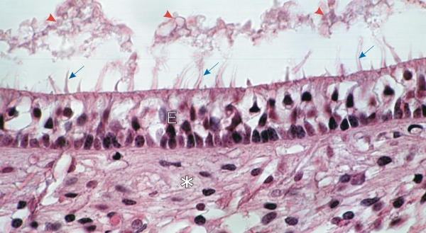
Section of the macula from the utricle of the ear of a monkey.

Sitting on a cushion of connective tissue and nerve fibres (*), the stratified epithelium (E) is composed of supporting cells and sensory hair cells. These sensory hair cells have tufts of microvilli or stereocilia (blue arrows) that plunge into a gelatinous layer mainly dissolved in this section. At the surface of this macula and gelatinous material (red arrowheads) there are miniscule crystalline bodies, or otoliths, which are difficult to identify in the present preparation.

Stain: H–­­E
Magnification: ×900


We'd very much appreciate receiving your comments and suggestions!We'd very much appreciate receiving your comments and suggestions!

Creative Commons Licence The text and images of this Histology Atlas, by Yves Clermont, Michael Lalli & Zsuzsanna Bencsath-Makkai, are licensed under a Creative Commons Attribution-Noncommercial-No Derivative Works 2.5 Canada Licence and cannot be modified without the written permission of the authors. Use of any text or images must carry an acknowledgement which includes a link to the original work.


HAweb/Zs B-M -> HAbase-generated on 2013-09-13