|
||
| 17. Ear | ||
| 1 2 3 4 5 6 7 8 9 10 11 |
| |||
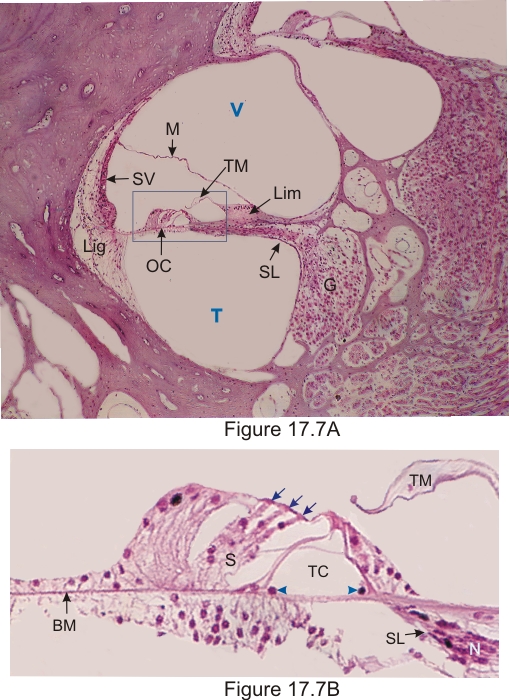 |
Figure 17.7A. Cochlea and cochlear duct.
The following elements are labelled:
The framed area is shown in Figure 17.7B. Figure 17.7B Organ of Corti and associated structures of a rodent. The following structures are labelled:
The tectorial membrane, condensed by the fixation, is artificially separated here from the sensory hair cells of the organ of Corti. The fine thread crossing the tunnel of Corti, in the direction of the hair cells, belongs to nerve fibers of the nerve (N) located in the osseous spiral lamina (SL) and reaching the spiral ganglion (see Figure 17.8). Stain: HE
|
||