 |
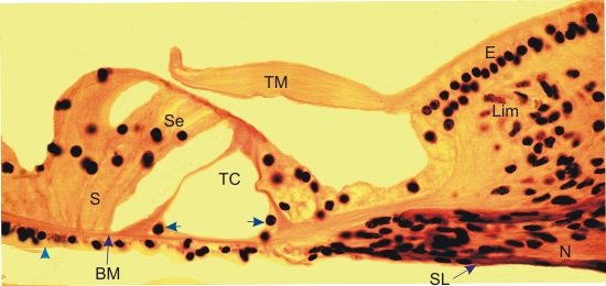
This figure shows the extremity of the osseous spiral lamina (SL) with a bundle of myelinated nerve fibres (N) in its core. On top of this bony lamina, the spiral limbus (Lim) is lined with a columnar epithelium (E) continuous with the tectorial membrane (TM). The latter, composed of a fibro-gelatinous substance overlies the epithelial cells of the organ of Corti.
The organ of Corti is composed of supporting cells (S) and sensory hair cells (Se). The triangular space in this organ, called the tunnel of Corti (TC), is delimited by pillar cells (arrows). Underlying this sensory epithelium, the thin basilar membrane (BM) is lined with a thin epithelium (arrowhead) facing the scala tympani.
The vibrations of the basilar membrane, originating from the middle ear, are transmitted to the hair cells whose apical microvilli are in contact with the tectorial membrane.
Stain: HE
Magnification: ×350
|