|
||
| 17. Ear | ||
| 1 2 3 4 5 6 7 8 9 10 11 |
| |||
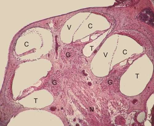 |
Longitudinal section of the cochlea of a guinea pig.
The following components of the cochlea are visible:
The details of the cochlear duct are shown in Figure 17.5. Stain: HE
|
||
|
||
| 17. Ear | ||
| 1 2 3 4 5 6 7 8 9 10 11 |
| |||
|
|
Longitudinal section of the cochlea of a guinea pig.
The following components of the cochlea are visible:
The details of the cochlear duct are shown in Figure 17.5. Stain: HE
|
||
![]() The text and images of this Histology Atlas, by Yves Clermont,
Michael Lalli & Zsuzsanna Bencsath-Makkai,
are licensed under a
Creative Commons Attribution-Noncommercial-No Derivative Works 2.5 Canada Licence
and cannot be modified without the written permission of the authors.
Use of any text or images must carry an acknowledgement which includes a link to the original work.
The text and images of this Histology Atlas, by Yves Clermont,
Michael Lalli & Zsuzsanna Bencsath-Makkai,
are licensed under a
Creative Commons Attribution-Noncommercial-No Derivative Works 2.5 Canada Licence
and cannot be modified without the written permission of the authors.
Use of any text or images must carry an acknowledgement which includes a link to the original work.