Contents Yves Clermont, Michael Lalli, Zsuzsanna Bencsath-Makkai
LIGHT-MICROSCOPIC HISTOLOGY ATLAS
     17. Ear
  1     2     3     4     5     6     7     8     9     10     11  

    17.8   Spiral ganglion Version française
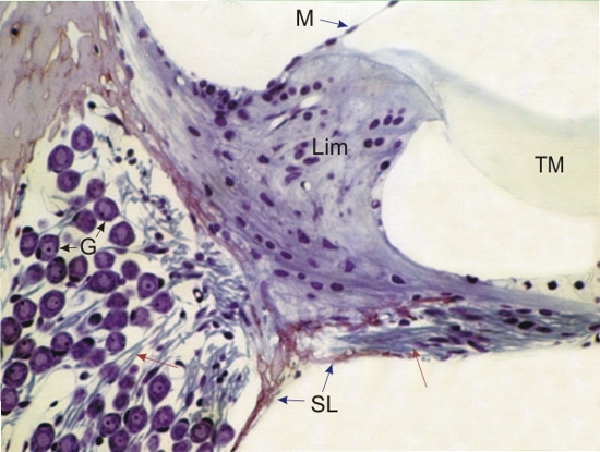
Section of the spiral ganglion located in the central axis of the cochlea, and of the proximal portion of the cochlear duct of a mouse.

The cells of the spiral ganglion (G) with their basophilic cytoplasm and spherical nucleus show their nerve fibres (red arrows) extending into the core of the osseous spiral lamina (SL) in the direction of the organ of Corti.

The following are also labelled:

  • Spiral limbus (Lim)
  • Tectorial membrane (TM)
  • Small segment of the vestibular membrane (M) of the cochlear duct

Stain: Toluidine blue
Magnification: ×900


We'd very much appreciate receiving your comments and suggestions!We'd very much appreciate receiving your comments and suggestions!

Creative Commons Licence The text and images of this Histology Atlas, by Yves Clermont, Michael Lalli & Zsuzsanna Bencsath-Makkai, are licensed under a Creative Commons Attribution-Noncommercial-No Derivative Works 2.5 Canada Licence and cannot be modified without the written permission of the authors. Use of any text or images must carry an acknowledgement which includes a link to the original work.


HAweb/Zs B-M -> HAbase-generated on 2013-09-13