|
||
| 17. Ear | ||
| 1 2 3 4 5 6 7 8 9 10 11 |
| |||
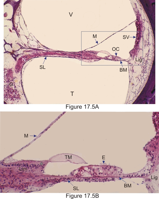 |
Figure 17.5A. Cochlea of a human ear.
This organ includes the spiral ganglion (G) with its nerve fibres (N) extending into the centre of the osseous spiral lamina (SL) in the direction of the organ of Corti (OC), the scala vestibuli (V), and the scala tympani (T). The triangular cochlear duct is delimited by the vestibular membrane (M), the stria vascularis (SV) and the organ of Corti (OC) sitting on the basilar membrane (BM). The spiral ligament (Lig) is identified. The framed area is shown at a higher magnification in Figure 17.5B. Figure 17.5B. Portion of the cochlear duct. The following structures are labelled: the vestibular membrane (M), the spiral limbus (Lim) lined with a columnar epithelium (*) connected to the tectorial membrane (TM) overlying the organ of Corti, which includes a sensory epithelium (E) sitting on a basilar membrane (BM). The basilar membrane bridges the tip of the spiral lamina (SL) and the spiral ligament (Lig) (see Figure 17.2). Stain: HE
|
||