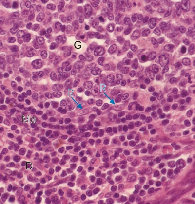
Sommaire Yves Clermont, Michael Lalli, Zsuzsanna Bencsath-Makkai
ATLAS D'HISTOLOGIE EN MICROSCOPIE OPTIQUE
     6. Organes Lymphatiques
  1     2     3     4     5     6     7     8     9     10     11     12     13     14     15     16     17     18     19     20     21     22     23     24     25  
  26     27     28     29     30     31     32     33     34     35     36     37     38  

    6.20   Ganglion lymphatique English version
Coupe du cortex d’un ganglion lymphatique.

Ce champ montre une portion du nodule avec son centre germinatif (G) et son manteau (Ma). Au bas de la photo sous le nodule on observe une zone de tissu lymphatique diffus (D).

Le centre germinatif contient de nombreux grands et moyens lymphocytes mais relativement peu de petits lymphatiques. Les noyaux allongés de deux cellules réticulaires sont indiqués (R). Le manteau (Ma) est composé de nombreux petits lymphocytes disposés en couches concentriques.

Coloration: H–­­É
Grossissement: ×600


We'd very much appreciate receiving your comments and suggestions!We'd very much appreciate receiving your comments and suggestions!

Creative Commons Licence The text and images of this Histology Atlas, by Yves Clermont, Michael Lalli & Zsuzsanna Bencsath-Makkai, are licensed under a Creative Commons Attribution-Noncommercial-No Derivative Works 2.5 Canada Licence and cannot be modified without the written permission of the authors. Use of any text or images must carry an acknowledgement which includes a link to the original work.


HAweb/Zs B-M -> HAbase-generated on 2013-09-13