Sommaire Yves Clermont, Michael Lalli, Zsuzsanna Bencsath-Makkai
ATLAS D'HISTOLOGIE EN MICROSCOPIE OPTIQUE
     6. Organes Lymphatiques
  1     2     3     4     5     6     7     8     9     10     11     12     13     14     15     16     17     18     19     20     21     22     23     24     25  
  26     27     28     29     30     31     32     33     34     35     36     37     38  
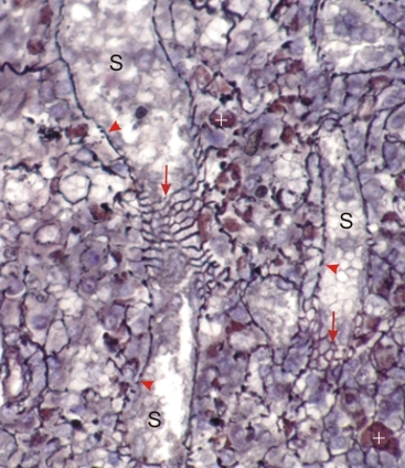
    6.37   Rate English version
Coupe de la rate d’un rongeur colorée à l’argent pour mettre en evidence, en noir, les fibres réticulaires.

Ce champ montre des réseaux de fibres épaisses autour des sinus veineux. Ce réseau argyrophile est vu de face (flèches) ou de profil (pointes de flèches).

Des macrophages de couleur brunâtre (+) sont distribués parmi les fibres réticulaires des cordons spléniques.

Coloration: Hématoxyline et Argent
Grossissement: ×750


We'd very much appreciate receiving your comments and suggestions!We'd very much appreciate receiving your comments and suggestions!

Creative Commons Licence The text and images of this Histology Atlas, by Yves Clermont, Michael Lalli & Zsuzsanna Bencsath-Makkai, are licensed under a Creative Commons Attribution-Noncommercial-No Derivative Works 2.5 Canada Licence and cannot be modified without the written permission of the authors. Use of any text or images must carry an acknowledgement which includes a link to the original work.


HAweb/Zs B-M -> HAbase-generated on 2013-09-13