Sommaire Yves Clermont, Michael Lalli, Zsuzsanna Bencsath-Makkai
ATLAS D'HISTOLOGIE EN MICROSCOPIE OPTIQUE
     6. Organes Lymphatiques
  1     2     3     4     5     6     7     8     9     10     11     12     13     14     15     16     17     18     19     20     21     22     23     24     25  
  26     27     28     29     30     31     32     33     34     35     36     37     38  
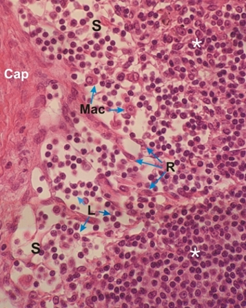
    6.17   Ganglion lymphatique English version
Coupe du cortex d’un ganglion lymphatique.

Ce champ montre un sinus sous-capsulaire (S) situé entre la capsule (Cap) et le tissu lymphatique diffus (*) du cortex.

Ce sinus est composé des éléments cellulaires suivants: les cellules réticulaires (R) qui forment un réseau de soutien; des lymphocytes (L); des macrophages (Mac).

Coloration: H–­­É
Grossissement: ×400


We'd very much appreciate receiving your comments and suggestions!We'd very much appreciate receiving your comments and suggestions!

Creative Commons Licence The text and images of this Histology Atlas, by Yves Clermont, Michael Lalli & Zsuzsanna Bencsath-Makkai, are licensed under a Creative Commons Attribution-Noncommercial-No Derivative Works 2.5 Canada Licence and cannot be modified without the written permission of the authors. Use of any text or images must carry an acknowledgement which includes a link to the original work.


HAweb/Zs B-M -> HAbase-generated on 2013-09-13