Contents Yves Clermont, Michael Lalli, Zsuzsanna Bencsath-Makkai
LIGHT-MICROSCOPIC HISTOLOGY ATLAS
     7. Blood and Bone Marrow
  1     2     3     4     5     6     7     8     9     10     11     12     13     14     15     16     17     18     19     20     21  

    7.11   Platelets Version française
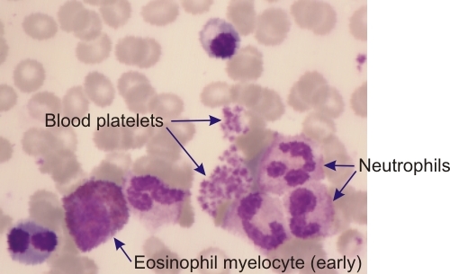
This field is a high magnification of a bone marrow smear.

Clusters of blood platelets are seen next to the neutrophils. An eosinophil myelocyte is also present nearby.

The blood platelets show a purple core and a pale acidophilic cortex.

Stain: Wright-Giemsa
Magnification: ×1000


We'd very much appreciate receiving your comments and suggestions!We'd very much appreciate receiving your comments and suggestions!

Creative Commons Licence The text and images of this Histology Atlas, by Yves Clermont, Michael Lalli & Zsuzsanna Bencsath-Makkai, are licensed under a Creative Commons Attribution-Noncommercial-No Derivative Works 2.5 Canada Licence and cannot be modified without the written permission of the authors. Use of any text or images must carry an acknowledgement which includes a link to the original work.


HAweb/Zs B-M -> HAbase-generated on 2013-09-13