Contents Yves Clermont, Michael Lalli, Zsuzsanna Bencsath-Makkai
LIGHT-MICROSCOPIC HISTOLOGY ATLAS
     7. Blood and Bone Marrow
  1     2     3     4     5     6     7     8     9     10     11     12     13     14     15     16     17     18     19     20     21  
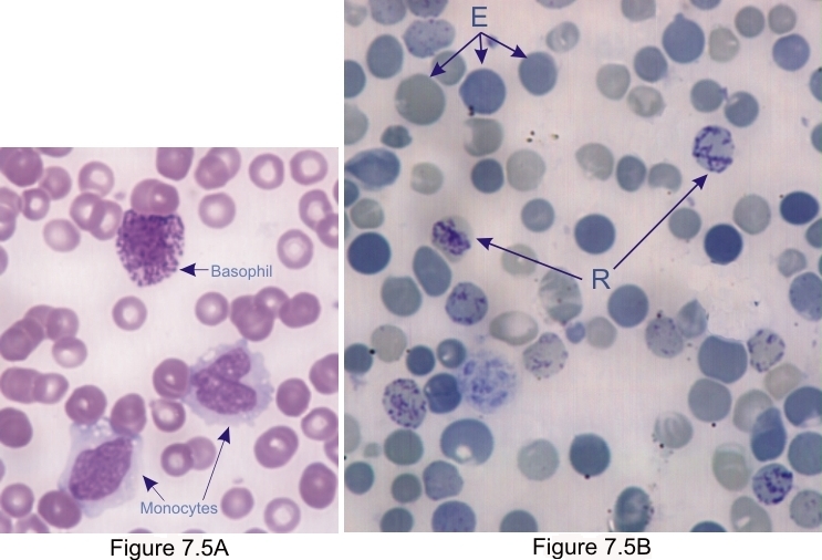
    7.5   Reticulocytes and leukocytes Version française
Figure 7.5A
  • Basophil
  • Monocytes

Figure 7.5B. Image of a blood smear fixed and stained with crezyl blue showing erythrocytes (E) in various shades of blue-green.

The smear also contains cells called reticulocytes (R), that is, erythrocytes containing basophilic and granulated networks. These networks are composed of precipitated ribosomes.

These reticulocytes, which are immature erythrocytes, compose 1% to 2% of the erythrocyte population. Higher percentages of these reticulocytes in the blood indicate an increased production of red blood cells in stimulated bone marrow under the influence of erythropoietin, which is produced in a hemorrhage.

Stain: Wright-Giemsa (7.5A); Crezyl blue (7.5B)


We'd very much appreciate receiving your comments and suggestions!We'd very much appreciate receiving your comments and suggestions!

Creative Commons Licence The text and images of this Histology Atlas, by Yves Clermont, Michael Lalli & Zsuzsanna Bencsath-Makkai, are licensed under a Creative Commons Attribution-Noncommercial-No Derivative Works 2.5 Canada Licence and cannot be modified without the written permission of the authors. Use of any text or images must carry an acknowledgement which includes a link to the original work.


HAweb/Zs B-M -> HAbase-generated on 2013-09-13