Contents Yves Clermont, Michael Lalli, Zsuzsanna Bencsath-Makkai
LIGHT-MICROSCOPIC HISTOLOGY ATLAS
     7. Blood and Bone Marrow
  1     2     3     4     5     6     7     8     9     10     11     12     13     14     15     16     17     18     19     20     21  

    7.10   Megakaryocyte Version française
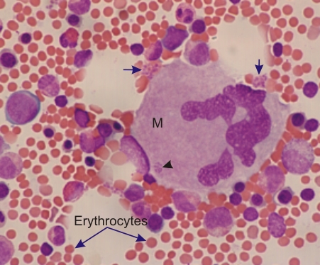
Bone marrow smear stained with the Wright-Giemsa method.

This field shows a megakaryocyte (M) surrounded by several hemopoietic cells, the precursors of erythrocytes and leukocytes.

The megakaryocyte shows a single large lobulated polyploid nucleus. This enormous nucleus is the result of several successive endomitoses.

The profuse cytoplasm is pale blue because of the ribosomes and purplish because of the fine specific azurophilic granules.

Small areas of the cytoplasm form clusters of blood platelets (arrowhead) and are seen at the surface of the megakaryocyte (arrows).

Stain: Wright-Giemsa
Magnification: ×600


We'd very much appreciate receiving your comments and suggestions!We'd very much appreciate receiving your comments and suggestions!

Creative Commons Licence The text and images of this Histology Atlas, by Yves Clermont, Michael Lalli & Zsuzsanna Bencsath-Makkai, are licensed under a Creative Commons Attribution-Noncommercial-No Derivative Works 2.5 Canada Licence and cannot be modified without the written permission of the authors. Use of any text or images must carry an acknowledgement which includes a link to the original work.


HAweb/Zs B-M -> HAbase-generated on 2013-09-13