|
||
| 7. Blood and Bone Marrow | ||
| 1 2 3 4 5 6 7 8 9 10 11 12 13 14 15 16 17 18 19 20 21 |
| |||
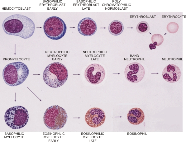 |
For more details on individual hemopoeietic cells see Table 7.2.
Coloured drawing of blood cell formation in bone marrow smears. This drawing, which is similar to that in Figure 7.8 should help in identifying the cells in figures 7.13 to 7.21 showing smears stained with Wright-Giemsa method. (Drawing done by Margo Oeltzschner) The chromatin of nuclei of the various cells is either stippled or flaky. The nucleoli are visible at early stages of differentiation. The cytoplasm presents various shades of blue and this basophilia is due to the ribonucleic acid of ribosomes. Cytoplasmic specific granules are azurophilic, pale eosinophilic or deeply acidophilic (orange in colour). The acidophilia of polychromatophilic erythroblasts, erythroblasts and erythrocytes is due to the presence of an increasing amount of hemoglobin. Lymphocytes, which are also produced in bone marrow and present in the smears, are not included in this drawing but are identified in the following smears.
|
||