Sommaire Yves Clermont, Michael Lalli, Zsuzsanna Bencsath-Makkai
ATLAS D'HISTOLOGIE EN MICROSCOPIE OPTIQUE
     7. Sang et Moelle Osseuse
  1     2     3     4     5     6     7     8     9     10     11     12     13     14     15     16     17     18     19     20     21  
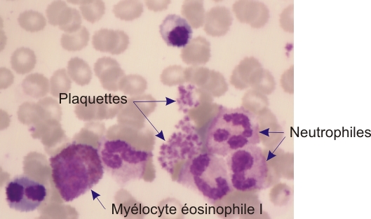
    7.11   Plaquettes English version
Photo au fort grossissement d’un frottis de moelle osseuse.

Ce champ montre des aggrégats de plaquettes sanguines parmi des neutrophiles en formation. Un myélocyte éosinophile est également présent à proximité.

Les plaquettes montrent un centre azurophile et un cortex légèrement acidophile.

Coloration: Wright-Giemsa
Grossissement: ×1000


We'd very much appreciate receiving your comments and suggestions!We'd very much appreciate receiving your comments and suggestions!

Creative Commons Licence The text and images of this Histology Atlas, by Yves Clermont, Michael Lalli & Zsuzsanna Bencsath-Makkai, are licensed under a Creative Commons Attribution-Noncommercial-No Derivative Works 2.5 Canada Licence and cannot be modified without the written permission of the authors. Use of any text or images must carry an acknowledgement which includes a link to the original work.


HAweb/Zs B-M -> HAbase-generated on 2013-09-13