|
||
| 7. Blood and Bone Marrow | ||
| 1 2 3 4 5 6 7 8 9 10 11 12 13 14 15 16 17 18 19 20 21 |
| |||
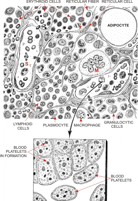 |
Drawing showing the composition of the red bone marrow (top) and the mode of formation of blood platelets (below) in a megakaryocyte (M).
The two venous sinuses (S) shown in the top diagram are the sites where newly formed erythrocytes and leukocytes migrate from the connective tissue to the blood circulation. The framework of this connective tissue is composed of stellate reticular cells associated with reticular fibres (collagen type III). Hemopoietic cells, which are involved in the production of erythrocytes and leukocytes, accumulate in the interstices between these supporting elements. Islands of erythroid, neutrophilic, eosinophilic and lymphoid cells are schematically illustrated in this drawing. Other cell types are also present in the bone marrow: adipocytes, monocytes, macrophages, plasma cells and megakaryocytes. Megakaryocytes with their polyploid nucleus are involved in the production of blood platelets. These platelets are often formed in the projections of megakaryocytes that enter into the lumen of a blood sinus. One such process, as seen with the electron microscope, is shown at the bottom of the drawing. The cytoplasm of this cell is partitioned by flattened cisternae that demarcate small areas of the cytoplam. These flattened cisternae fuse and liberate, at the surface of the cell, discoid bodies or platelets (from 1 to 3 µm in diameter), which circulate in the blood. The platelets contain the following elements: specific azurophilic granules, mitochondria, ribosomes, contractile filaments, microtubules and glycogen particles. These blood platelets may agglutinate to patch holes in the walls of damaged vessels or contribute to the coagulation of blood.
|
||