Sommaire Yves Clermont, Michael Lalli, Zsuzsanna Bencsath-Makkai
ATLAS D'HISTOLOGIE EN MICROSCOPIE OPTIQUE
     7. Sang et Moelle Osseuse
  1     2     3     4     5     6     7     8     9     10     11     12     13     14     15     16     17     18     19     20     21  

    7.21   Moelle osseuse English version
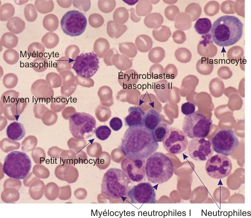
Frottis de moelle osseuse montrant les cellules étiquetées suivantes:
  • un myélocyte basophile
  • érythroblastes basophiles II
  • myélocytes neutrophiles I
  • deux neutrophiles
  • petits et moyen lymphocytes

Un plasmocyte est également présent. Il montre un petit noyau condensé et un cytoplasme basophile abundant. Une zone peri-nucléaire golgienne pâle est aussi bien distincte.

Coloration: Wright-Giemsa
Grossissement: ×1400


We'd very much appreciate receiving your comments and suggestions!We'd very much appreciate receiving your comments and suggestions!

Creative Commons Licence The text and images of this Histology Atlas, by Yves Clermont, Michael Lalli & Zsuzsanna Bencsath-Makkai, are licensed under a Creative Commons Attribution-Noncommercial-No Derivative Works 2.5 Canada Licence and cannot be modified without the written permission of the authors. Use of any text or images must carry an acknowledgement which includes a link to the original work.


HAweb/Zs B-M -> HAbase-generated on 2013-09-13