|
||
| 5. Vaisseaux | ||
| 1 2 3 4 5 6 7 8 9 10 11 12 13 14 15 16 17 18 19 20 21 22 23 24 25 | ||
| 26 27 28 29 30 31 32 33 34 35 |
| |||
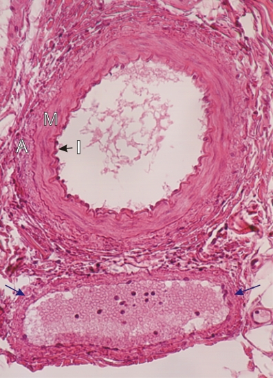 |
Coupes transversales dune artère (en haut) et de la veine qui laccompagne (en bas).
Lartère montre les trois tuniques habituelles: lintima (I), la média (M) et ladventice (A). La paroi plus mince de la veine montre du tissu conjonctif (flèches bleues) mais les cellules musculaires lisses ne sont pas identifiables ici. Coloration: HÉ
|
||