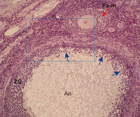
Sommaire Yves Clermont, Michael Lalli, Zsuzsanna Bencsath-Makkai
ATLAS D'HISTOLOGIE EN MICROSCOPIE OPTIQUE
     16. Appareil Génital Féminin
  1     2     3     4     5     6     7     8     9     10     11     12     13     14     15     16     17     18     19     20     21     22     23     24     25  
  26     27     28     29     30     31     32     33     34     35     36     37     38     39     40     41     42     43     44     45     46     47  

    16.13   Atrésie folliculaire English version
Coupe du cortex ovarien d’un singe. Le champs encadré est présenté à un plus fort grossissement dans la figure 16.14.

À proximité d’un follicule primaire multilaminaire normal (Pa-m) on note la présence d’un gros follicule secondaire à antrum (An) qui montre les signes précurseurs d’une atrésie folliculaire.

Cette régression se manifeste par une désorganisation de la zona granulosa (ZG) et la présence de cellules folliculaires ou granuleuses en dégénérescence avec des noyaux condensés ou en pycnose {flèches).

Coloration: H–­­É
Grossissement: ×250


We'd very much appreciate receiving your comments and suggestions!We'd very much appreciate receiving your comments and suggestions!

Creative Commons Licence The text and images of this Histology Atlas, by Yves Clermont, Michael Lalli & Zsuzsanna Bencsath-Makkai, are licensed under a Creative Commons Attribution-Noncommercial-No Derivative Works 2.5 Canada Licence and cannot be modified without the written permission of the authors. Use of any text or images must carry an acknowledgement which includes a link to the original work.


HAweb/Zs B-M -> HAbase-generated on 2013-09-13