Sommaire Yves Clermont, Michael Lalli, Zsuzsanna Bencsath-Makkai
ATLAS D'HISTOLOGIE EN MICROSCOPIE OPTIQUE
     16. Appareil Génital Féminin
  1     2     3     4     5     6     7     8     9     10     11     12     13     14     15     16     17     18     19     20     21     22     23     24     25  
  26     27     28     29     30     31     32     33     34     35     36     37     38     39     40     41     42     43     44     45     46     47  
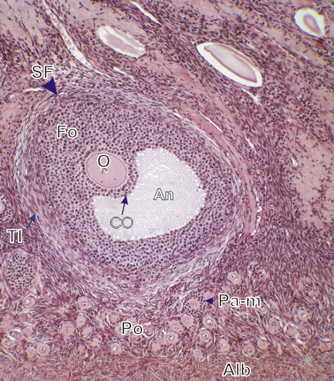
    16.10   Cortex ovarien English version
Coupe d’un follicule secondaire ou antral (SF).

L’antrum (An), rempli de liquide folliculaire, est entouré de cellules folliculeuses (Fo) ou granuleuses.

Certaines de ces cellules qui entourent l’oocyte (O) forment le cumulus oophorus (CO).

Dans le stroma la thèque interne ou theca interna (TI) est nettement visible à la surface de ce follicules secondaires.

On observe égalemnent, entre ce follicule et l’albuginée ou tunica albuginea (Alb), de nombreux follicules primordiaux (Po) et quelques follicules primaires (Pa-m).

Coloration: H–­­É
Grossissement: ×250


We'd very much appreciate receiving your comments and suggestions!We'd very much appreciate receiving your comments and suggestions!

Creative Commons Licence The text and images of this Histology Atlas, by Yves Clermont, Michael Lalli & Zsuzsanna Bencsath-Makkai, are licensed under a Creative Commons Attribution-Noncommercial-No Derivative Works 2.5 Canada Licence and cannot be modified without the written permission of the authors. Use of any text or images must carry an acknowledgement which includes a link to the original work.


HAweb/Zs B-M -> HAbase-generated on 2013-09-13