Sommaire Yves Clermont, Michael Lalli, Zsuzsanna Bencsath-Makkai
ATLAS D'HISTOLOGIE EN MICROSCOPIE OPTIQUE
     1. Épithéliums
  1     2     3     4     5     6     7     8     9     10     11     12     13     14     15     16     17     18     19     20     21     22     23     24  
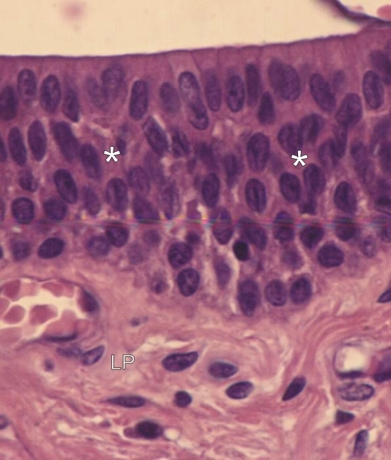
    1.21   Épithélium stratifié prismatique de l’urètre English version
Épithélium stratifié prismatique d’un urètre humain.

Cet épithélium montre une couche de cellules prismatiques à la surface et, au dessous, quelques couches superposées de cellules polyhédriques ou allongées (*).

Dans cet épithélium, seulement les cellules basales sont en contact avec le tissu conjonctif de la lamina propria (LP).

Cet épithélium est donc réellement stratifié.

Coloration: H–­­É
Grossissement: ×600


We'd very much appreciate receiving your comments and suggestions!We'd very much appreciate receiving your comments and suggestions!

Creative Commons Licence The text and images of this Histology Atlas, by Yves Clermont, Michael Lalli & Zsuzsanna Bencsath-Makkai, are licensed under a Creative Commons Attribution-Noncommercial-No Derivative Works 2.5 Canada Licence and cannot be modified without the written permission of the authors. Use of any text or images must carry an acknowledgement which includes a link to the original work.


HAweb/Zs B-M -> HAbase-generated on 2013-09-13