Sommaire Yves Clermont, Michael Lalli, Zsuzsanna Bencsath-Makkai
ATLAS D'HISTOLOGIE EN MICROSCOPIE OPTIQUE
     8. Cartilage et Os
  1     2     3     4     5     6     7     8     9     10     11     12     13     14     15     16     17     18     19     20     21     22     23     24     25  
  26     27     28     29     30     31  

    8.29   Spicules mixtes English version
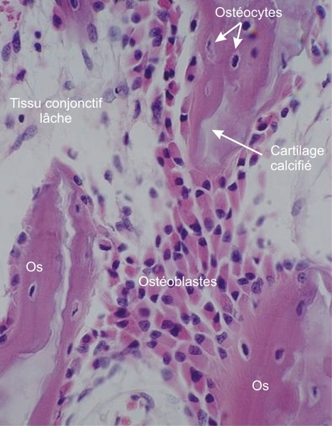
Spicules mixtes sous un plateau épiphysaire d’un jeune chien.

Ce champ montre, en vue de face, une couche d’ostéoblastes très chromophiles associée à trois portions de spicules mixtes. Les ostéocytes de l’os acidophile sont logés dans leurs logettes.

Du cartilage calcifié basophile est présent dans le centre des spicules.

Coloration: H–­­É
Grossissement: ×400


We'd very much appreciate receiving your comments and suggestions!We'd very much appreciate receiving your comments and suggestions!

Creative Commons Licence The text and images of this Histology Atlas, by Yves Clermont, Michael Lalli & Zsuzsanna Bencsath-Makkai, are licensed under a Creative Commons Attribution-Noncommercial-No Derivative Works 2.5 Canada Licence and cannot be modified without the written permission of the authors. Use of any text or images must carry an acknowledgement which includes a link to the original work.


HAweb/Zs B-M -> HAbase-generated on 2013-09-13