Contents Yves Clermont, Michael Lalli, Zsuzsanna Bencsath-Makkai
LIGHT-MICROSCOPIC HISTOLOGY ATLAS
     4. Nerve Tissue
  1     2     3     4     5     6     7     8     9     10     11     12     13     14     15     16     17     18     19     20     21     22     23     24     25  
  26     27     28     29     30     31     32     33     34     35  

    4.7   Ganglion Version française
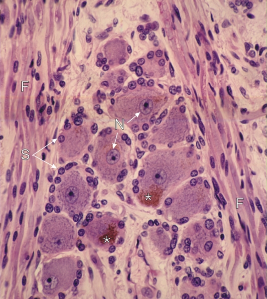
Section of a spinal ganglion.

Several ganglion cells show a spherical nucleus (N) with a large central basophilic nucleolus. The surrounding cytoplasm shows basophilic Nissl bodies. Some of these cells show a cluster of brownish-red granules (*) in the perikaryon. These are lipofuscin granules which derive from lysosomes.

The small cells with a spherical nucleus which closely surround the ganglion cells are satellite cells (S), a class of neuroglia.

On each side of this group of ganglion cells, poorly myelinated nerve fibres (F) are seen in longitudinal or oblique sections.

Note that in this preparation the Golgi bodies are not intensely stained.

Stain: H–­­E
Magnification: ×900


We'd very much appreciate receiving your comments and suggestions!We'd very much appreciate receiving your comments and suggestions!

Creative Commons Licence The text and images of this Histology Atlas, by Yves Clermont, Michael Lalli & Zsuzsanna Bencsath-Makkai, are licensed under a Creative Commons Attribution-Noncommercial-No Derivative Works 2.5 Canada Licence and cannot be modified without the written permission of the authors. Use of any text or images must carry an acknowledgement which includes a link to the original work.


HAweb/Zs B-M -> HAbase-generated on 2013-09-13